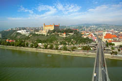
 The first Hungarian university (Studium Generale Quinqueecclesiensis) was founded by King Louis the Great in Pécs in 1367 , with Pope Urban V's permission. The medical was one of the faculties of the university which followed the four-faculty European pattern. Although the university had to suspend its operation after a few decades, due to the unfortunate historical events, on the basis of the discovered documents there was constantly some level of medical training in Pécs. The revival of University of Pécs was a consequence of the Treaty of Trianon. The Hungarian Royal Elizabeth University that launched education in Bratislavain 1914 was obliged to leave the ancient coronation city in 1919 and the university arrived to Pécs by Mecsek in autumn 1923, after nearly half a decade of wandering in Budapest.
The theoretical departments of Royal Elizabeth University ahad not been ready by the foundation of the medical faculty in Bratislava in 1918, therefore the education in Bratislava started only in the upper years. The theoretical departments were founded in the capital only after the 1919 escape from Bratislava, at first together with the other university escaped, namely with the Franz Joseph University in Cluj-Napoca which later got to Szeged.
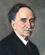
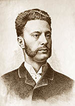 Professor Mihály Lenhossék (1863-1937) was teaching anatomy in the capital for consolidated years of the two universities escaped in the school year 1920/21 in the classroom and laboratories of the No. 1. Department of Anatomy in Budapest (at 58. Tűzoltó Street). Lenhossék gave his lectures entitled "Regular Anatomy with Topographical Relations". The practices called ""Anatomical and Histological Practices were also held (managed) by Lenhossék for first-year students every day throughout the afternoon and on Saturday mornings in the dissecting room and in the histological laboratory of the Department. In the first school year, 1920/21 in Budapest 391 students enrolled to the Medical Faculty of Elizabeth University in the first semester and 629 in the second. The number of students increased to 1204 in the next school year, as well as in the last one, 1922/23 in the capital. The change in the last two school years in Budapest was that the courses were - partly - held in the veterinary college. The number of medical students was 885 in the first semester in Pécs, 680 in the second, 510 in the third, 398 in the fourth and from that time the number of medical students varied between 300 and 400; there was a further decrease from the mid 1930s when their number was between 200 and 250.
 The Department of Anatomy was founded in Pécs in 1923, when The Hungarian Royal Elizabeth University was moved to Pécs.The department was placed in the former sports hall (at 5. Dischka Győző utca). The leadership of the Department and the teaching of anatomy were solved for a long time by Zsigmond Tóth's (1872-1950) appointment as head of the department, since professor Tóth had led the Department until 1944, age of 72. The Department provided theoretical and practical education for an average of 60 or 70 students each year, then from 1942 to the end of the war for a maximum of 100 students. In the spacious dissecting rooms the students attended the practical lessons together, usually managed by an assistant lecturer and one or two trainees or demonstrators. Due to the cadaver supply that was lower than today, first-year students could preparate only limbs and they could only study the anatomy of body cavities in the scope of so-called situs demonstrations, standing on stands around the cadaver being preparated by the assistant lecturer. Whole cadavers could be preparated only by second-year students under an incomparably smaller teaching management than today. In the histological practices about 50 or 60 slides, all of them stained with haematoxylin-eosin were presented for only 20 students at the same time, because only one room and 21 practical microscopes were available.
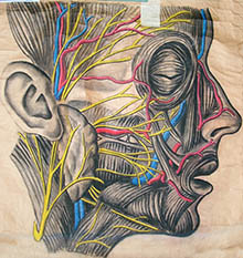
In accordance with the much narrower educational framework than today, there was a small number of teachers: there was only one associate professor, one assistant professor and two trainees paid, completed by unpaid trainees and demonstrators. There was available equipment to research mainly in the field of descriptive anatomy, histology and embryology in the department. Since Zsigmond Tóth, the first director of the Department was primarily keen on increasing the number of illustrative demonstration materials: the work of the department, besides strict educational work, mostly consisted of making preparations and educational figures. A significant part of the current figure storage and museum material is due to the efficiency of these works. Professor Tóth not only gave these lectures, but also managed dissecting practices in the large, undivided dissecting rooms by the help of associate and assistant lecturers, trainees and demonstrators. Altogether 92 students worked in the department under his management. Later on many of them had an outstanding academic and other professional career; for instance, Mihály Bodosi, Kornél Dvorszky, Tihamér Gy. Karlinger, Kálmán Kisfaludy, Lajos Kollár, Dezső Kollár, Margit Mittag, Ernő Mózsa, Károly Röhlich, Pál Schwartz, Elemér Scipiades, Júlia Szanathy and Nándor Than.
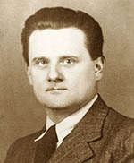 After Professor Tóth's retirement the management and anatomy education of the department was taken by associate professor Vereby (Röhlich) Károly (1900-1945) who was an extraordinary university professor passed away at a young age. In 1944, in accordance with the opportunities and the limited research needs Vereby had the non-educational rooms of the department modernized. He set up the museum room and the preparation room. After the conversion the central part of the department was a large laboratory attached to a staff room, a library, a smaller laboratory and a fairly tight experimental animal operating room. In the latter only the most necessary and fairly primitive equipment was available. However, the equipment of the photo laboratory attached to the practical room downstairs was good enough. Besides Professor Tóth's anthropological and embryological work, Károly Vereby carried out an extremely high-standard and valuable research in histology and embryology as an assistant and associate lecturer, which he developed into significant experimental studies also in foreign relations in bone regeneration particularly in his last years.
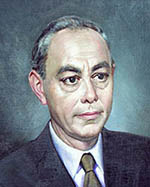
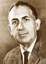 Professor Vereby, died tragically in the summer of 1945, was not given the chance to provide the development of the department he had just taken over. The about half-year interregnum following the director of the department's death barely served the keeping of the situation. Géza Mansfeld, the Dean of the Faculty entrusted Miklós Melczer, professor of dermatology with the education of anatomy, under the management of Professor Béla Entz. As a result of Béla Entz Rector's intention (1945-1946), the management of Hungarian Royal Elizabeth University and that of Medical Faculty decided to invite János Szentágothai (1912-1994) who was in U.S. military captivity to be Head of Department and to continue the education of anatomy. Szentágothai became a private professor in 1946 March and an academic professor in 1948. Although Kálmán Lissák Rector proposed Szentágothai to become regular university professor three times between 1947 and 1949, Szentágothai not only received no appointment, but his payment and all his benefits were revoked in 1949 by the Scientific Secretariat of the Council of Ministers despite of his acclaimed work, written together with Ferenc Kiss, Atlas of the Anatomy of the Human Body had been published half a decade before. In the following years, the atlas had a total of 82 editions in 32 languages. Finally on September 15th, 1951 Szentágothai became a professor- he was still only 39 years old.
The educational work of the department was carried out in accordance with the old educational forms, the courses were still held at Dischka Street, but with higher number of students. Significant difficulties occurred due to the location of the department and also because in the years following the war there was only one medical doctor at the department in addition to the director: Tibor Kiss, later surgeon professor, who was persuaded by Szentágothai in 1947 to move to Pécs as an associate professor, leaving his assistant lecturer position in Budapest. Thus he became the youngest associate professor in the country at the age of 25. The other part of the assistance solely consisted of enthusiastic medical students and demonstrators. Béla Flerkó, who already was a demonstrator at the department lead by professor Tóth, became a prize-winning demonstrator as a medical student on September 1st, 1947.
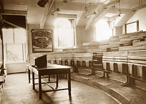 |
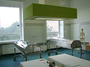
| A Classroom of the Former Department of Anatomy |
A new educational (the so-called group) system had been introduced in the school year 1948/49 at the Medical Faculty of University of Pécs (the name of which had changed). Those five or six groups of students, who worked and were educated together in the only dissecting room available, disturbed each other's education. Therefore, through the internal construction line, professor Szentágothai provided better conditions for the education of increasing number of students in the dissecting room, primarily with dividing the dissecting room into six smaller premises by the help of partitions, each one with the capacity of one student group. A further increase in the level of anatomical education was the result of the improvement of the cadaver supply by the help of county councils, so for each first- and second-year group of students one cadaver was available for dissection. At this time many preparations of Kiss-Szentágothai atlas were already being made in Pécs.
There were further changes in the name and structure of the university on February 1st, 1951, which resulted in the separation of the Medical Faculty and the Faculty of Law, as well as that of Teacher Training College. From this time, the former Medical Faculty became the Medical University of Pécs (POTE).
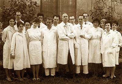 |
| Staff of the Department in 1962 |
Filling the gradually increasing number of positions with qualified doctors educated in the department resulted in the establishment of the teaching staff, which is twice as much in number than 20 years before in that assistant professors, senior lecturers, assistant lecturers and trainees teach anatomy and histology for ten groups of first-year and ten groups of second-year students. A place for research also had to be provided for the increasing number of the staff, due to the dual job of the department. The non-educational part of the department was rebuilt gradually from 1949, which began with the enlargement of the experimental animal operating room and the establishment of a small histological laboratory, to the detriment of a large staff room. The southern front in the central part of the department got its recent layout that time. The enlarged animal operating room got equipment according to the experimental morphological work trend that had been increasingly growing. Two years later the northern front was reconstructed practically, when one oversized laboratory was converted to two laboratories and an office, and the unutilized foreground of the previous large laboratory became an animal dissecting room attached to the operating room and a library. Thus the previous library was also successfully converted for a better laboratory use. Later on the department also got the living room next to the photo laboratory on the ground floor. Another quite large room became successfully free after the photo laboratory had been moved to the premise separated from the preparatory classroom upstairs. The isotope part of the Department was created after the practical division of these two rooms.
 The outlined internal reconstructions ensured properly the placement of the increasing number of the staff and the conditions of the research that meanwhile torn into several fields, but could only moderately relieve the crowdedness caused by the originally narrow frameworks. The problem was radically solved in 1954 by the set up of the new Department of Histology and Embryology. The Department was founded in the rooms belonging to the former Otorhinolaryngology Acut Care Unit (at 33. Sallai Street) that became free by the establishment of Otorhinolaryngology Clinic. The department was to be independent, but after the unsuccessful contest for department leadership, professor Szentágothai was entrusted with the leadership of this department as well. The two histological practical rooms and with the change of completely obsolete microscopes, the microscope park increased to fourty pieces enabled the parallel education of two groups of students. Besides the completion of the basic series (stained with haematoxylin-eosin), the slide set for practical lessons was increased by preparation series stained and impregnated by different methods so that the set of the department consists of 140 different preparation series, especially the neurohistological part of which is very versatile.
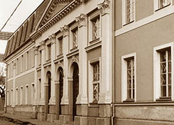 |
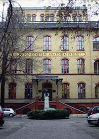
Frontage of the old Anatomy Institute
(Dischka Gy. street) |
Besides two educational rooms three new research laboratories and a new small animal operating room (for rats and guinea-pigs) were located in the Department of Histology and Embryology, that due to the proper equipment of the laboratories enabled the neuroendocrine researches, a main work trend that had been developed since the 1950s, to be moved from Department of Anatomy to the Department of Histology and Embryology. This research especially required a suitable animal research facility that could be established by the help of the Section of Medical Sciences of the Hungarian Academy of Sciences (HAS). Besides, this enabled the creation of an own strain of Wistar rats in the department. Laboratories that became empty after a part of the neuroendocrinological research work got to the Department of Histology and Embryology provided space for the experimental neuroembryological group that emerged in the meantime. After professor Szentágothai's departure to Budapest, the operations of dogs and cats that had previously been popular practically stopped, the Department of Anatomy somewhat relieved its lack of space by converting the animal operating room into a histochemical laboratory. The electron microscope that was given to the department by the Section of Biological Sciences of HAS (Hungarian Academy of Sciences) in 1965 got place in the laboratory built in the backyard of the Department of Anatomy. During the reconstruction of a part of the anatomy building, the dissection area got renovated and the animal research facility was somewhat extended.
In Szentágothai's time, the department launched an intensive research work. The first research direction derived from that Professor Szentágothai moved his previous researches in synapse degeneration to his new workplace. Thereafter and by the introduction of the stereotaxic method, the department became an important methodical centre of the modern approach to functional neuroanatomy and that of its boundaries, from where not only several inland, but some foreign scientific institutes also took over the stereotaxic methods. These methods together with synapse-degeneration histological techniques made synaptology the first primary field of research of the department that targets the study of the connection between the structure and function of axon terminals. It was professor Szentágothai who developed the most modern experimental, morphological methods of these researches (namely synapse degeneration, "isolated spinal cord and cerebral cortex"- method) and thus he clarified many elemental neurophysiological, synaptological and synapse histochemical problems with his colleagues in connection with the spinal cord, the cerebellum, and the cerebral cortex.. The stereotaxic methods were also the methodical basis for nerve pathway researches on the detection of neuronal organisation of some major neural pathways and reflex arches. The second biggest research field of the department, the study of neuroendocrine control rooted in those efforts of some young, new trainees and demonstrators in the department who had started their research in the late 40s, that determine the tissue reactions of certain endocrine glands and that of their hormonal appendices through quantitative histological methods (nuclear variation statistics, quantitative differential cell-identification etc.). After these efforts the attention inevitably turned to the tissue reactions of troph hormones of the pituitary, then to the effects of diencephalon and that of the whole central nervous system (CVS). The third field of scientific research in the department was experimental neuroembryology. Herewith the Department intended to evoke the scientific method called experimental embryology that was used almost only by Károly Vereby in the country. Similarly to the previous field, the difficulty this time came from that in the situation of the early 50s young people in the department interested in the topic had to learn the necessary methods completely self-taught, by themselves.
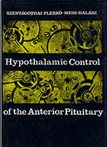
Szentágothai established extended international connections with outstanding foreign, primarily American, English, French and Italian research laboratories. Due to these connections and Szentágothai's national and international prestige, the teaching staff and researchers at the Department got into the exceptional situation that even from behind the "iron curtain", they could go to a generally one-year study tour in the 1960s and 1970s. The group got into international scientific forefront in a short time with topics of the structure of the spinal cord and that of cerebellum, also with the CVS control of hypophysis. Among others, the monography entitled Hypothalamic Control of the Anterior Pituitary with more editions dates back to this period, which is one of the fundamental works describing the physiology and control mechanisms of hypophysis. There were several modern methods developed and introduced. Halász-knife, introduced in 1968, became one of the most successful tools of contemporary modern brain research. As a result of György Sétáló's study tour in Denver, the first European immunohistochemical laboratory was launched in our Department in 1968.
Professor Szentágothai established an excellent school in Pécs. In this period, Szentágothai's students became heads of the departments of anatomy at medical universities one after another in the country. Szentágothai himself, then Miklós Réthelyi became head of No.1. Department of Anatomy in Budapest, Béla Halász became head of No.2. Department of Anatomy and the head of Department in Debrecen became Professor György Székely. The director of Department of Anatomy at University of Veterinary Medicine was also chosen from this institute, in the person of Ferenc Hajós. Later in the 90s, the Department in Szeged was also led by an anatomist from Pécs called Zsolt Liposits. Besides leading the departments of anatomy, numerous students of Szentágothai's school became internationally recognized professors, researchers or extraordinary clinicians at home and foreign departments, such as József Hámori, György Illei, András Gömöri, Béla Török, Tihamér Gy. Karlinger, Tibor Kiss, Miklós Palkovits, Miklós Réthelyi, Antal Salamon, Béla Török, László Jaszmann, Péter Petrusz, Károly Strazniczky, Béla Kosaras - just to mention some of them.
 After Szentágothai's departure in 1963, Béla Flerkó took over the direction of the Department who became a professor at the age of 39 like Szentágothai and was head of department for almost thirty years. The new theoretical block of POTE was opened in 1970 in 12. Szigeti Street. The Department of Anatomy and Institute of Histology moved here from Dischka Győző Street (where the building of Dental Clinic currently is) and from Ferencesek Street. The Department established two histological practical rooms and five completely separated dissecting rooms in the new building. In addition, experimental laboratories also had substantially bigger space. The Department set up an own laboratory with electron microscope and an animal research facility. This time Anatomy and Histology were four-semester courses with four autopsy practices, two histological practices and with two to four lectures per week. The lectures of 250-270 students per year were primarily given by Béla Flerkó - with the contribution of professors Béla Mess and György Székely, the practises for ten groups per year were held by associate professor Ágnes Donhoffer; furthermore, assistant lecturers called Károly Strazniczky, György Sétáló, Lajos Tima, Gyula Lázár, Béla Kosaras and István Lengvári. Béla Flerkó was one of those professors at the Department who also fulfilled the Rector's office (1979-1985). Mihály Lenhossék was Rector at Royal Hungarian University in the school year 1914-1915 while Zsigmond Tóth was Rector in the school year 1940-1941 at Hungarian Royal Elizabeth University. The medical education in English at POTE started in 1983, during Béla Flerkó's second period as a Rector. Lajos Tima, a member of the Department had been secretary for English education from the beginning until 2004.
There were several educational reforms introduced at POTE in the 1970s and 1980s. Practically there were barely two sequential academic years with same structures at POTE in this period. Anatomy teaching lasted sometimes three, sometimes four semesters. There was "integrated education" for years with physiology and biochemistry education. The number of classes was also changed several times.
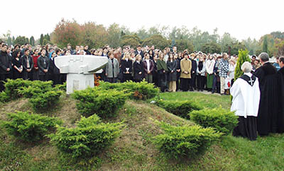 |
| Funerary Commemoration |
Uniquely in the country, the course of Anatomy, Histology and Embryology has been taught through three semesters in Pécs since 1986, but the total number of classes is practically equivalent to that of other medical faculties. Our cadaver supply in education became critical in the early '90s. Until then the cadavers used in education had been selected from the dead without relatives in twilight homes. The changes that had come along with the changing political system had cut these sources. A new system could be established primarily due to professor Judit Horváth's sacrificial work, based on voluntary offers of people who decided to serve medical education with their body after their death. The initiation was given significant help by setting aside the churches and the media. There has been a separate, well-kept parcel placed for the cadavers in the cemetery and each year the Department give thanks to its offerers on the memorial on All Souls' Day in the presence of numerous students, teachers and that of representatives of churches. Thanks to them, the Department can even cover the educational need of the increased number of students.
The Department continued its international-level research. The researches were still performed in the fields of neuroendocrinology and in neurohistology. Several modern methods were developed or introduced. For the first time in the country, professor Valér Csernus set up a steroid RIA laboratory in 1973 that still has an important role in the research work of the Department, with its own developed technique and antibodies of great quality. Tamás Görcs and Zsolt Liposits developed the silver and nickel-intensification procedure in immunohistochemistry on light and electron microscopic level, which resulted in a significant increase in the sensitivity of the method. Taking the advantages of a short study tour in the Netherlands, Sándor Vígh introduced a modern in vitro bioassay method - a superfusional and perfusional technology - in the Department in 1986. The method has been developed further by other researchers of the Institute. As a result, the Department became an international centre of this method. Researchers of our Department set up, sustain and give professional advice to superfusional laboratories in many renowned research institutes all over the world, primarily in the United States and Germany.
Members of the Department occupied prominent places not only in home, but in international scientific public life. Among researchers who had been educated at the Department János Szentágothai, Béla Flerkó, Béla Halász, György Székely and József Hámory have become HAS members so far. János Szentágothai was Chairman of HAS for two periods; Béla Halász was Vice Chairman of HAS. Researchers at our Department took long-term jobs at renowned foreign universities and research institutes, due to the outstanding reputation of our Department.
 Péter Petrusz is Director of Department of Cell and Developmental Biology at University of North Carolina, Károly Strazniczky was head of Department of Anatomy at The Flinders University of South Australia (Adelaide), Béla Kosaras works at Department of Neurology at Harvard University, István Merchenthaler led a research laboratory at Women's Health Research Institute of Wyeth Ayerst company, he currently works at a research laboratory of NIH (National Institutes of Health) in Chapel Hill; Sándor Vígh is Professor at Department of Anatomy at Tulane University (New Orleans).
In accordance with the provisions, Béla Flerkó, age 65, gave the Department leadership to Professor György Sétáló in 1992 who led the Institute until 2002. Professor Flerkó had participated actively in the work of the Department as a research professor until his death in 2003. Professor György Sétáló still participates actively in the educational work of the Department as a professor emeritus.
Based on a political decision, the University of Pécs (Pécsi Tudományegyetem, PTE) was found on January 1st, 2000, into which most institutions of higher education in the region merged. Thus POTE ended its operation and became one of the ten faculties of PTE called Medical School. Simultaneously with the change, the name of the Department also changed for a better distinction from similar departments of other faculties of PTE: it became Department of Human Anatomy of Medical School of University of Pécs.
The Director of the Institute was professor Valér Csernus between 2002-2012. In the year 2002 the name of the Department changed back to Department of Anatomy that meets the traditions and the international customs in a better way. The credit-based training had been introduced. The Department underwent significant development in the subsequent years. The need for change derived not only from the introduction of the credit-based system, but from the sudden increase in the number of students. Instead of the previous 180 to 200 Hungarian and 40 to 60 English students, in three degree programmes (general medicine, dentistry, pharmacy) of the first two years (first and second) in 2007 we had already taught 500 Hungarian, 430 English and 410 German students altogether. The increasing number of students also required the development of infrastructure. The Department established two other dissecting rooms and another histology room in 2006. There are modern air-conditioners and lighting system in the dissecting rooms. There are twenty-six modern microscopes available for the students in each of the three histology rooms. Instead of the slideshow there is a computer projection system in each histology room, as well as a microscope camera connected to a widescreen television; both facilitate education significantly.
 |
| A New Dissecting Room |
The subject structure changed according to the modern educational principles and without the substantive change of the number of classes or that of the syllabus. Students currently learn macroscopic anatomy, histology and embryology as specific subjects through two-two semesters, then they learn neuroanatomy through one semester ending in a complex final exam. The Department introduced the system of organized mid-semester exams. Our students write tests by the help of computer projected images each semester, the result is a part of the final mark. The system of mid-semester exams increased the students' learning spirit and also improved the exam result. Some clinician lecturers are also invited to give a lecture. High-standard anatomy and histology lectures filled with clinical experiences are popular among students and the authentic medical experiences increased the popularity of the otherwise monotonous course. The Department announces numerous optional courses besides the obligatory ones. The most popular ones are the dissection courses, during which students dissect cadavers also used in obligatory practical classes with more thoroughness, due to more time is available. It not only increases the students' chances of getting a cadaver, but it also gives free range to more explanations during the obligatory classes. The two-dimensional anatomy course is also popular that enables students to compare the images from modern imaging processes to their three-dimensional anatomy experiences, involving clinician lecturers.
The teaching staff of the Department has been sustaining an educational and information system (an-server.pote.hu) on the Internet for approximately ten years to facilitate students' learning and informing. The websites of about seven thousand files, available all over the world, not only provide the current information for students, but also includes the entire curriculum of histology, interactive and subtitled photos of cadavers and the demonstrative materials of the lectures. Animations and video-recordings also facilitate learning.
Unfortunately, the research work is considerably difficult due to the educational burdens that have been significantly increased (the teaching staff of the Department has to teach 296 hours each week). Nevertheless, the Department is still one of those which produce the most scientific evidences at the university, as a result of serious efforts. The teaching staff of the Department published about 131 books and chapters in the last thirty years,
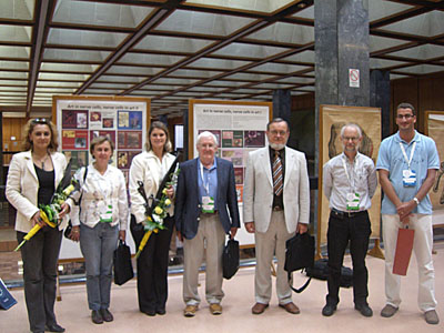 |
Organizers of CECE and
Management of ESCE in the AulaA |
they also wrote 886 articles for scientific journals mostly in English for internationally renowned medical journals. The total impact factor of these articles is over two thousand, the number of citations is more than twelve thousand. Out of the 20 full-time and 8 part-time lecturers of the Department, 20 have Ph.D degree, 6 have DSc. degree and 13 have Dr.Habil degree. Lecturers of the Department are members and board members of several national and international scientific societies and they are also editorial members of many high-level international scientific journals. Professor Valér Csernus is full member of the German Academy of Sciences (Deutsche Akademie der Naturforscher Leopoldina). Members of the Department also participate successfully in home and international scientific tenders.
There are regularly six to eight current, previously won benefit tenders that help research work with tens of millions of forints. Apart from our experimental methods continued traditionally - such as stereotaxic surgeries, immunohistochemistry, on the level of light and electronmicroscopy, pathway researches, RIA, superfusion - there have been new modern methods introduced; for instance, molecular biological methods (RT-PCR, blotting, gene-silencing), methods of tissue culture and those that study the behaviour of animals, and there have been laboratories set up according to these methods. Our lecturers participate in the work of national as well as international organisations and corporations. Many of them are members of the public body of HAS.The Department is in fruitful cooperation with several national and foreign research institutes from Chile to Japan. Ten members of the Department had gained Ph.D degree between 2002 and 2011. The Department has 15 Ph.D students in 2011. Our TDK (Undergraduate Research Sociaty) students also help our educational work and do very well with their scientific achievements in national and international conferences. In 2002 the Department organized the symposium called"Biological rhythmic processes. The function of biological clock in the living world". The Department also organised the 11th Congress of the Hungarian Society for Neuroscience Research, as well as the 13rd Congress of the Hungarian Society of Anatomy in 2005 and in 2010 it organised the 25th Congress of the European Society for Comparative Endocrinology (CECE-25).
In 2011 the Department was involved in the arrangement of the common Conference of Hungarian Society for Physiology, Hungarian Society for Anatomy and Hungarian Society for Pharmacology.
The teaching staff participates actively in the public life of the faculty and that of the university. Professor Gyula Lázár had been Academic Vice Rector for over a period. Professor Valér Csernus was Vice Dean for Education over two periods and also Vice Dean for General Affairs, Education and Science for over one period. The teaching staff participates in the work and are members of numerous academic and faculty committees. Among others professor Gyula Lázár was Chairman of the Habilitation Committee of the Faculty, professor Valér Csernus was Chairman of the Curriculum Committee, professor Judit Horváth was Chairwoman of the Educational Committee, is now the Chairwomen of the Credit Transfer Committee, professor István Lengvári was Chairman of K+F Commission, professor Zoltán Rékási is Secretary of the Doctoral Committee. Professor Béla Mess was advisor of the German Programme Committee.
The director of the Institute has been professor Dóra Reglődi since January of 2013. The Vice-Chairman is professor Judit Horváth.
Here you can find the current lecturers and staff of the Department.